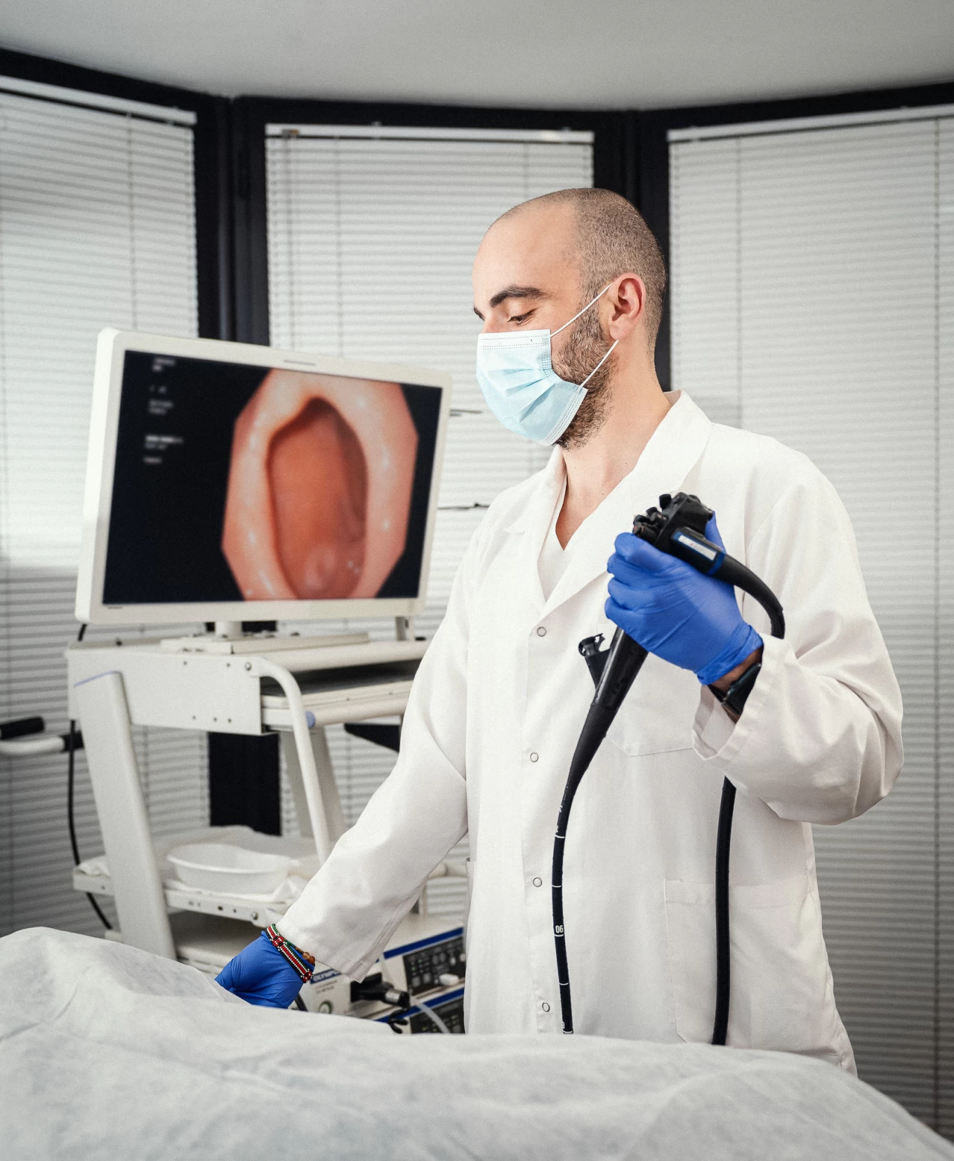
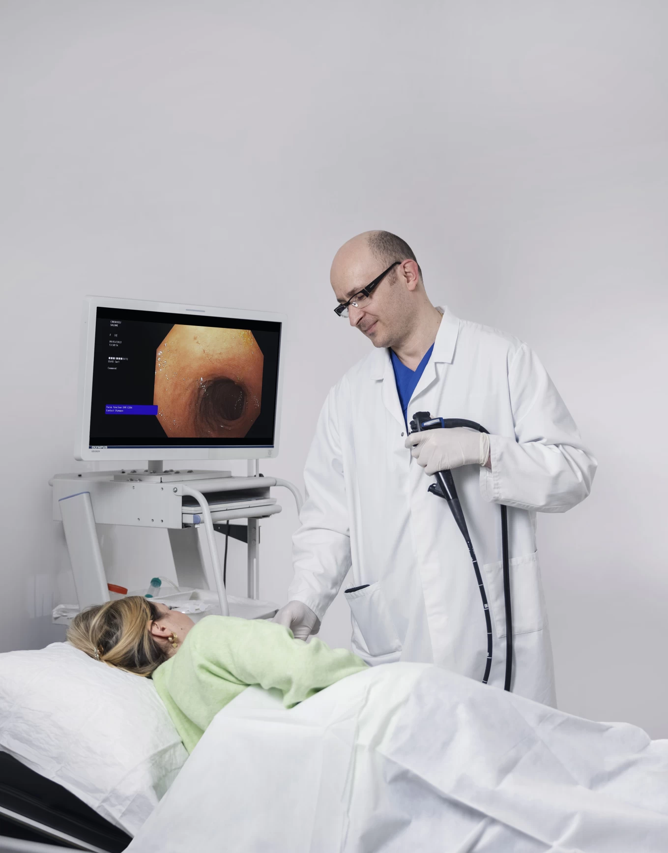
Endoscopy Department at the Vera Branch
The department conducts:
- Endoscopic examination of the esophagus, stomach, duodenum, and large intestine, as well as biopsy-based detection of pyloric Helicobacter infections.
- Balloon dilation of strictures caused by achalasia, burns, and anastomoses of the esophagus.
- Early detection of stomach and intestinal cancer.
- Determination of the endoscopic and morphological patterns of various forms of colitis.
Examinations are performed under local anesthesia as well as venous sedation.
განყოფილებაში დამონტაჟებულია ფირმა OLYMPUS - ის ულტრათანამედროვე ციფრული ენდოსკოპიური აპარატი Evis Exera III, რომელიც გამოირჩევა გამოსახულების მაღალი ხარისხით, რის შედეგადაც ხდება პათოლოგიური პროცესის უფრო მკაფიოდ შესწავლა. წინა მოდელის აპარატებთან შედარებით Evis Exera III-ის ორფოკუსიანი ვიდეოკამერა მაქსიმალურად უახლოვდება გამოსაკვლევ მიდამოს. ახლოფოკუსიანი ვიზუალიზაციისა და ფერების შეცვლის ფუნქციით ხდება არსებული პათოლოგიის ადრეულ სტადიაზე დიაგნოსტირება. Evis Exera III წარმოადგენს ოქროს სტანდარტს კიბოსწინარე დაავადებების დიაგნოსტიკასა და სიმსივნური წარმონაქმნების ვიზუალიზაციაში. NBI ფუნქციით შესაძლებელია ქსოვილის კაპილარული ქსელის მიკროსკოპული ვიზუალიზაცია და სხვადასხვა დაავადებების დიფერენცირება. სხვა ენდოსკოპიურ სისტემებთან შედარებით გაუმჯობესებულია ენდოსკოპიური ულტრაბგერითი ფუნქცია, რაც გულისხმობს უშუალოდ ენდოსკოპიური პროცედურის დროს ამა თუ იმ უბნის ულტრაბგერით კვლევას.
The department is equipped with the state-of-the-art digital endoscopic device Evis Exera III by OLYMPUS, which is distinguished by high image quality, allowing for a more detailed study of pathological processes. Compared to previous models, the Evis Exera III’s dual-focus video camera brings the area of examination closer. With the new focus visualization and color change function, early-stage diagnosis of existing pathology is possible. Evis Exera III represents the gold standard in the diagnosis of precancerous conditions and the visualization of neoplastic formations. The NBI function allows for microscopic visualization of the tissue’s capillary network and the differentiation of various diseases. Compared to other endoscopic systems, the endoscopic ultrasound function has been improved, enabling ultrasound examination of specific areas during the endoscopic procedure.


